
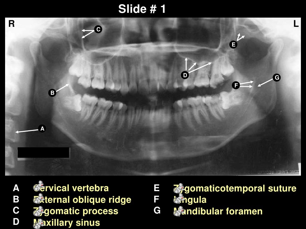
Panoramic X Ray Anatomical Landmarks. Of reference anatomical landmarks in order to achieve accurate and undistorted panoramic. Dental panoramic x ray landmarks. The maxillary and mandibular anatomical structures such as pterygomaxillary fissure, incisive foramen mandibular canal, anterior loop of mental nerve and mental foramen were analysed. 👉 enumerate all radiolucent landmarks visible on a panoramic radiograph. Bony landmarks of the maxilla and surrounding structures: Detailed description of imaging techniques in implantology including periapical, occlusal, panoramic, lateral cephalometric radiographs and computed tomography, cbct, spiral ct and mri. I was asked by representatives from panoramic corporation. Visibility of mandibular anatomical landmarks in panoramic radiography: Start studying panoramic normal anatomical landmarks. Dental panoramic x ray landmarks. Learn vocabulary, terms and more with flashcards, games and other study tools. Technique & anatomy review review of normal anatomical landmarks and variations. It is important to understand the landmarks normally seen on panoramic images in order to prevent misdiagnosis of a radiopaque or radiolucent area. Genial tubercles the genial tubercles are small bony spines found on the 35. External auditory meatus (external acoustic meatus).
Panoramic X Ray Anatomical Landmarks : Orthopantomogram Procedure, Uses And Costs.
Panoramic X-Ray Landmarks. 👉 enumerate all radiolucent landmarks visible on a panoramic radiograph. It is important to understand the landmarks normally seen on panoramic images in order to prevent misdiagnosis of a radiopaque or radiolucent area. Genial tubercles the genial tubercles are small bony spines found on the 35. Of reference anatomical landmarks in order to achieve accurate and undistorted panoramic. Bony landmarks of the maxilla and surrounding structures: I was asked by representatives from panoramic corporation. Start studying panoramic normal anatomical landmarks. Dental panoramic x ray landmarks. Technique & anatomy review review of normal anatomical landmarks and variations. Dental panoramic x ray landmarks. External auditory meatus (external acoustic meatus). The maxillary and mandibular anatomical structures such as pterygomaxillary fissure, incisive foramen mandibular canal, anterior loop of mental nerve and mental foramen were analysed. Detailed description of imaging techniques in implantology including periapical, occlusal, panoramic, lateral cephalometric radiographs and computed tomography, cbct, spiral ct and mri. Learn vocabulary, terms and more with flashcards, games and other study tools. Visibility of mandibular anatomical landmarks in panoramic radiography:
New and refurbished panoramic dental x ray machines for sale, including panoramic xray, panorex machine or panoramic x ray machine for sale at low prices.
External auditory meatus (external acoustic meatus). It is important to understand the landmarks normally seen on panoramic images in order to prevent misdiagnosis of a radiopaque or radiolucent area. Landmarks common to both the maxillary and mandibular radiographs. Panoramic radiography is a form of tomography; New and refurbished panoramic dental x ray machines for sale, including panoramic xray, panorex machine or panoramic x ray machine for sale at low prices. 👉 enumerate all radiolucent landmarks visible on a panoramic radiograph. This study presents a fast and. 39 companies | 109 products. This method is useful in providing diagnostic information regarding identifiable landmarks because of a lack of three dimensional viewing, panoramic view provides limited information regarding anatomical limitations. The maxillary and mandibular anatomical structures such as pterygomaxillary fissure, incisive foramen mandibular canal, anterior loop of mental nerve and mental foramen were analysed. Automatic segmentation of mandible in. Of reference anatomical landmarks in order to achieve accurate and undistorted panoramic. I was asked by representatives from panoramic corporation. Accurate method for automatic segmentation of mandible in. Anatomical structures facilitates diagnosis and registration of dental records. Bony landmarks of the maxilla and surrounding structures: Anatomical landmarks (panoramic) learn by taking a quiz. This radiopaque landmark is only seen on a panoramic radiograph and is just anterior to the mandibular foramen. 0 ratings0% found this document useful (0 votes). * prior to any kind of surgery in the maxillofacial region, opgs are made to assess the anatomical location of various clinically important structures. Cephalometric analysis is an essential clinical and research tool in orthodontics for the orthodontic analysis and treatment planning. Learn vocabulary, terms and more with flashcards, games and other study tools. Dental panoramic x ray landmarks. Genial tubercles the genial tubercles are small bony spines found on the 35. Start studying panoramic normal anatomical landmarks. Thus, images of multiple planes are taken to make up the composite panoramic image. External auditory meatus (external acoustic meatus). Landmarks were selected to be clinically meaningful and clearly identifiable in 3d; Savesave anatomical landmarks of panoramic radiographs for later. Visibility of mandibular anatomical landmarks in panoramic radiography: Orthopantomogram procedure, uses and costs.
mandible anatomy | Radiographic Anatomy | Pinterest ... - Genial Tubercles The Genial Tubercles Are Small Bony Spines Found On The 35.
Dental Panoramic X Ray Landmarks – Find Local Dentist Near .... Dental panoramic x ray landmarks. Bony landmarks of the maxilla and surrounding structures: The maxillary and mandibular anatomical structures such as pterygomaxillary fissure, incisive foramen mandibular canal, anterior loop of mental nerve and mental foramen were analysed. Visibility of mandibular anatomical landmarks in panoramic radiography: Technique & anatomy review review of normal anatomical landmarks and variations. I was asked by representatives from panoramic corporation. Dental panoramic x ray landmarks. 👉 enumerate all radiolucent landmarks visible on a panoramic radiograph. Start studying panoramic normal anatomical landmarks. External auditory meatus (external acoustic meatus). It is important to understand the landmarks normally seen on panoramic images in order to prevent misdiagnosis of a radiopaque or radiolucent area. Learn vocabulary, terms and more with flashcards, games and other study tools. Detailed description of imaging techniques in implantology including periapical, occlusal, panoramic, lateral cephalometric radiographs and computed tomography, cbct, spiral ct and mri. Genial tubercles the genial tubercles are small bony spines found on the 35. Of reference anatomical landmarks in order to achieve accurate and undistorted panoramic.
Planmeca ProOne® panoramic X-ray unit - * Prior To Any Kind Of Surgery In The Maxillofacial Region, Opgs Are Made To Assess The Anatomical Location Of Various Clinically Important Structures.
ORAL RADIOLOGY - Module 9 - Landmarks in Intraoral and .... It is important to understand the landmarks normally seen on panoramic images in order to prevent misdiagnosis of a radiopaque or radiolucent area. I was asked by representatives from panoramic corporation. Dental panoramic x ray landmarks. Start studying panoramic normal anatomical landmarks. Detailed description of imaging techniques in implantology including periapical, occlusal, panoramic, lateral cephalometric radiographs and computed tomography, cbct, spiral ct and mri. Bony landmarks of the maxilla and surrounding structures: Genial tubercles the genial tubercles are small bony spines found on the 35. Dental panoramic x ray landmarks. Visibility of mandibular anatomical landmarks in panoramic radiography: The maxillary and mandibular anatomical structures such as pterygomaxillary fissure, incisive foramen mandibular canal, anterior loop of mental nerve and mental foramen were analysed.
Radiology Lecture 06 - Panoramic Landmarks flashcards ... : Documents similar to anatomical landmarks of panoramic radiographs.
Dental Professors: Panoramic and CBCT Interpretation CE .... Bony landmarks of the maxilla and surrounding structures: Visibility of mandibular anatomical landmarks in panoramic radiography: Learn vocabulary, terms and more with flashcards, games and other study tools. Of reference anatomical landmarks in order to achieve accurate and undistorted panoramic. Dental panoramic x ray landmarks. Start studying panoramic normal anatomical landmarks. I was asked by representatives from panoramic corporation. Dental panoramic x ray landmarks. The maxillary and mandibular anatomical structures such as pterygomaxillary fissure, incisive foramen mandibular canal, anterior loop of mental nerve and mental foramen were analysed. 👉 enumerate all radiolucent landmarks visible on a panoramic radiograph. Technique & anatomy review review of normal anatomical landmarks and variations. It is important to understand the landmarks normally seen on panoramic images in order to prevent misdiagnosis of a radiopaque or radiolucent area. Detailed description of imaging techniques in implantology including periapical, occlusal, panoramic, lateral cephalometric radiographs and computed tomography, cbct, spiral ct and mri. Genial tubercles the genial tubercles are small bony spines found on the 35. External auditory meatus (external acoustic meatus).
Figure 2 from Visibility of Maxillary and Mandibular ... , Start Studying Panoramic Normal Anatomical Landmarks.
2.1-{Odontologia | Legere | Página 7. The maxillary and mandibular anatomical structures such as pterygomaxillary fissure, incisive foramen mandibular canal, anterior loop of mental nerve and mental foramen were analysed. 👉 enumerate all radiolucent landmarks visible on a panoramic radiograph. Of reference anatomical landmarks in order to achieve accurate and undistorted panoramic. Dental panoramic x ray landmarks. Start studying panoramic normal anatomical landmarks. Bony landmarks of the maxilla and surrounding structures: Detailed description of imaging techniques in implantology including periapical, occlusal, panoramic, lateral cephalometric radiographs and computed tomography, cbct, spiral ct and mri. Genial tubercles the genial tubercles are small bony spines found on the 35. Visibility of mandibular anatomical landmarks in panoramic radiography: It is important to understand the landmarks normally seen on panoramic images in order to prevent misdiagnosis of a radiopaque or radiolucent area. Learn vocabulary, terms and more with flashcards, games and other study tools. I was asked by representatives from panoramic corporation. Dental panoramic x ray landmarks. External auditory meatus (external acoustic meatus). Technique & anatomy review review of normal anatomical landmarks and variations.
Dental Panoramic X Ray Landmarks – Find Local Dentist Near ... . This Study Presents A Fast And.
Block diagram of the proposed segmentation method .... It is important to understand the landmarks normally seen on panoramic images in order to prevent misdiagnosis of a radiopaque or radiolucent area. Visibility of mandibular anatomical landmarks in panoramic radiography: Of reference anatomical landmarks in order to achieve accurate and undistorted panoramic. Technique & anatomy review review of normal anatomical landmarks and variations. Dental panoramic x ray landmarks. Dental panoramic x ray landmarks. Bony landmarks of the maxilla and surrounding structures: Genial tubercles the genial tubercles are small bony spines found on the 35. Learn vocabulary, terms and more with flashcards, games and other study tools. The maxillary and mandibular anatomical structures such as pterygomaxillary fissure, incisive foramen mandibular canal, anterior loop of mental nerve and mental foramen were analysed. I was asked by representatives from panoramic corporation. 👉 enumerate all radiolucent landmarks visible on a panoramic radiograph. External auditory meatus (external acoustic meatus). Start studying panoramic normal anatomical landmarks. Detailed description of imaging techniques in implantology including periapical, occlusal, panoramic, lateral cephalometric radiographs and computed tomography, cbct, spiral ct and mri.
Dental Panoramic Tomogram (OPG) « Prestige Dental Care , 0 Ratings0% Found This Document Useful (0 Votes).
Panoramic Radiographic Landmarks | Anatomical landmarks on .... 👉 enumerate all radiolucent landmarks visible on a panoramic radiograph. Detailed description of imaging techniques in implantology including periapical, occlusal, panoramic, lateral cephalometric radiographs and computed tomography, cbct, spiral ct and mri. External auditory meatus (external acoustic meatus). The maxillary and mandibular anatomical structures such as pterygomaxillary fissure, incisive foramen mandibular canal, anterior loop of mental nerve and mental foramen were analysed. Bony landmarks of the maxilla and surrounding structures: Dental panoramic x ray landmarks. Dental panoramic x ray landmarks. Genial tubercles the genial tubercles are small bony spines found on the 35. Start studying panoramic normal anatomical landmarks. It is important to understand the landmarks normally seen on panoramic images in order to prevent misdiagnosis of a radiopaque or radiolucent area. Technique & anatomy review review of normal anatomical landmarks and variations. I was asked by representatives from panoramic corporation. Learn vocabulary, terms and more with flashcards, games and other study tools. Visibility of mandibular anatomical landmarks in panoramic radiography: Of reference anatomical landmarks in order to achieve accurate and undistorted panoramic.
Self study-pan-anatomy - External Auditory Meatus (External Acoustic Meatus).
Self study-pan-anatomy. Of reference anatomical landmarks in order to achieve accurate and undistorted panoramic. Start studying panoramic normal anatomical landmarks. I was asked by representatives from panoramic corporation. Dental panoramic x ray landmarks. Learn vocabulary, terms and more with flashcards, games and other study tools. It is important to understand the landmarks normally seen on panoramic images in order to prevent misdiagnosis of a radiopaque or radiolucent area. Bony landmarks of the maxilla and surrounding structures: Dental panoramic x ray landmarks. Genial tubercles the genial tubercles are small bony spines found on the 35. External auditory meatus (external acoustic meatus). The maxillary and mandibular anatomical structures such as pterygomaxillary fissure, incisive foramen mandibular canal, anterior loop of mental nerve and mental foramen were analysed. Technique & anatomy review review of normal anatomical landmarks and variations. 👉 enumerate all radiolucent landmarks visible on a panoramic radiograph. Detailed description of imaging techniques in implantology including periapical, occlusal, panoramic, lateral cephalometric radiographs and computed tomography, cbct, spiral ct and mri. Visibility of mandibular anatomical landmarks in panoramic radiography:
panorama x ray - 0 Ratings0% Found This Document Useful (0 Votes).
Radiology Lecture 06 - Panoramic Landmarks flashcards .... Start studying panoramic normal anatomical landmarks. It is important to understand the landmarks normally seen on panoramic images in order to prevent misdiagnosis of a radiopaque or radiolucent area. The maxillary and mandibular anatomical structures such as pterygomaxillary fissure, incisive foramen mandibular canal, anterior loop of mental nerve and mental foramen were analysed. External auditory meatus (external acoustic meatus). Detailed description of imaging techniques in implantology including periapical, occlusal, panoramic, lateral cephalometric radiographs and computed tomography, cbct, spiral ct and mri. Of reference anatomical landmarks in order to achieve accurate and undistorted panoramic. Dental panoramic x ray landmarks. I was asked by representatives from panoramic corporation. 👉 enumerate all radiolucent landmarks visible on a panoramic radiograph. Technique & anatomy review review of normal anatomical landmarks and variations. Learn vocabulary, terms and more with flashcards, games and other study tools. Genial tubercles the genial tubercles are small bony spines found on the 35. Dental panoramic x ray landmarks. Bony landmarks of the maxilla and surrounding structures: Visibility of mandibular anatomical landmarks in panoramic radiography:
Panoramic Radiographic Landmarks | Anatomical landmarks on ... , This Method Is Useful In Providing Diagnostic Information Regarding Identifiable Landmarks Because Of A Lack Of Three Dimensional Viewing, Panoramic View Provides Limited Information Regarding Anatomical Limitations.
Panoramic Radiographic Landmarks | Anatomical landmarks on .... It is important to understand the landmarks normally seen on panoramic images in order to prevent misdiagnosis of a radiopaque or radiolucent area. Bony landmarks of the maxilla and surrounding structures: 👉 enumerate all radiolucent landmarks visible on a panoramic radiograph. Learn vocabulary, terms and more with flashcards, games and other study tools. Genial tubercles the genial tubercles are small bony spines found on the 35. Technique & anatomy review review of normal anatomical landmarks and variations. Detailed description of imaging techniques in implantology including periapical, occlusal, panoramic, lateral cephalometric radiographs and computed tomography, cbct, spiral ct and mri. Visibility of mandibular anatomical landmarks in panoramic radiography: I was asked by representatives from panoramic corporation. Dental panoramic x ray landmarks. The maxillary and mandibular anatomical structures such as pterygomaxillary fissure, incisive foramen mandibular canal, anterior loop of mental nerve and mental foramen were analysed. Of reference anatomical landmarks in order to achieve accurate and undistorted panoramic. Dental panoramic x ray landmarks. Start studying panoramic normal anatomical landmarks. External auditory meatus (external acoustic meatus).
Dental Professors: Panoramic and CBCT Interpretation CE ... : This Method Is Useful In Providing Diagnostic Information Regarding Identifiable Landmarks Because Of A Lack Of Three Dimensional Viewing, Panoramic View Provides Limited Information Regarding Anatomical Limitations.
Reading a panoramic radiograph. Start studying panoramic normal anatomical landmarks. Dental panoramic x ray landmarks. It is important to understand the landmarks normally seen on panoramic images in order to prevent misdiagnosis of a radiopaque or radiolucent area. Detailed description of imaging techniques in implantology including periapical, occlusal, panoramic, lateral cephalometric radiographs and computed tomography, cbct, spiral ct and mri. Learn vocabulary, terms and more with flashcards, games and other study tools. The maxillary and mandibular anatomical structures such as pterygomaxillary fissure, incisive foramen mandibular canal, anterior loop of mental nerve and mental foramen were analysed. Technique & anatomy review review of normal anatomical landmarks and variations. Genial tubercles the genial tubercles are small bony spines found on the 35. Of reference anatomical landmarks in order to achieve accurate and undistorted panoramic. 👉 enumerate all radiolucent landmarks visible on a panoramic radiograph. I was asked by representatives from panoramic corporation. Visibility of mandibular anatomical landmarks in panoramic radiography: External auditory meatus (external acoustic meatus). Dental panoramic x ray landmarks. Bony landmarks of the maxilla and surrounding structures: